Topic
Imaging using a variety of modalities, including electron microscopy, atomic force microscopy, optical microscopy, X-ray and neutron imaging, ultrasonic techniques, focused ion beam techniques, and more, as well as diverse methods for performing image analysis.
Target audience
Scientists, Ph.D.’s and post-docs working with different imaging techniques and image analysis methods. Anyone who is interested in learning about the latest developments in imaging and image analyses.
Objectives
The series of Empa Topical Days on Imaging and Image Analysis, an event of the Research Focus Area ‘Health and Performance’, offers scientists, both from the ETH domain and from other public/private institutions, a comprehensive platform to stay updated on the latest developments and share experiences in the fields of imaging and image analysis.
Kindly register by May 7, 2025
Program
08:30 Robert Zboray, Center for X-ray Analytics, Empa
Welcome & opening remarks
08:40 Ludovico SIlvestri, European Laboratory for Non-linear Spectroscopy (LENS), University of Florence
Large-scale high-resolution imaging of multiple molecular targets with light-sheet fluorescence microscopy
09:20 Kiarash Tajbakhsh, Center for X-ray Analytics, Empa
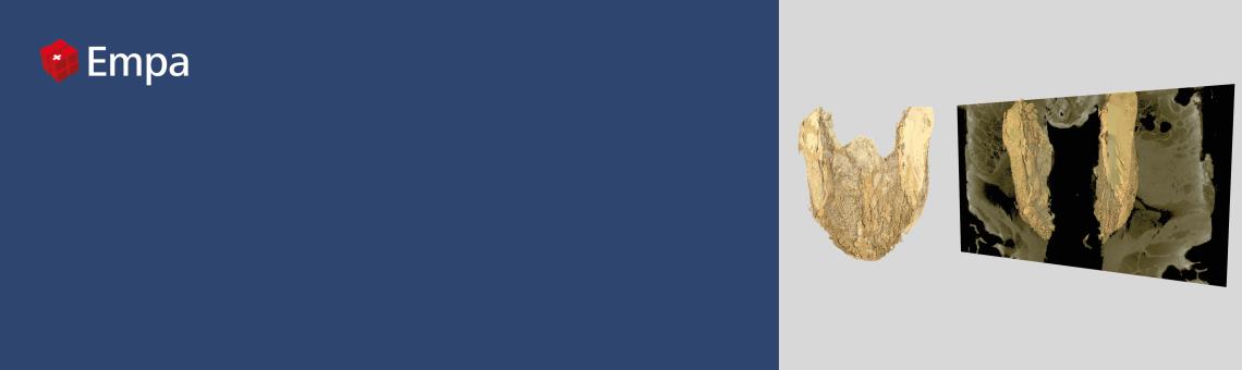
Thyroid neoplasm pathology from micro-anatomy to molecular signatures using X-ray imaging
10:00 Jingjing Jiang, Biomedical Optics Research Laboratory, Department of Neonatology,
University Hospital Zurich
Time domain near infrared optical tomography for clinical applications
10:40 Coffee break
11:00 Viktor Kis, 3DHISTECH, Budapest
Transforming pathology to 3D – Latest developments of μCT imaging by 3DHISTECH –
The Digital Pathology Company
11:40 Pavel Trtik, Applied Materials Group, Laboratory for Neutron Scattering and Imaging, PSI
Quasi-coherent assortment of neutron imaging activities at PSI – from ‘moderately useful’ to ‘quite useless
though rather interesting’ ones
12:20 Lunch break
13:20 Marianne Liebi, Laboratory for X-ray characterization of materials, EPF Lausanne & Structure and
Mechanics of Advanced Materials Group, Laboratory for Condensed Matter, Center for Photon Science, PSI
Imaging with X-ray scattering contrast for the characterization of hierarchical material structure
14:00 Sebastian Habermann, Laboratory Nanomaterials in Health, Department Materials meet Life, Empa
Colorful Insights: Pinpointing Biomolecules with Emissive Nanoparticles in Electron Microscopy
14:40 Coffee break
15:00 Rachele Butti & Vittorio Montanelli, Electron Microscopy Center, Empa
Crystal Orientation Mapping of All-Solid-State Battery Cathodes via 4D-STEM
Understanding Gold Nanoparticle Formation in Ionic Liquids via VT In Situ LP-STEM.
15:40 Andres Velasquez Parra, Department of Water Resources and Drinking Water, Eawag
Understanding solute transport and mixing in unsaturated porous media using 3D X-ray uCT
16:20 Closing